Histopathology of the Human Lens
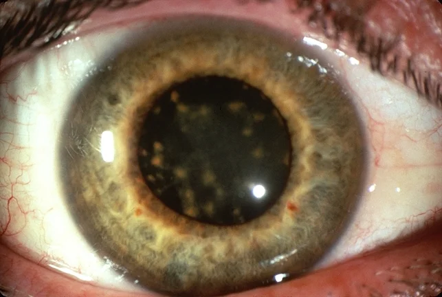
The human lens is positioned in the anterior 1/3 of the globe and provides about 1/3 of the focusing power of the eye. The lens contributes to morbidity through cataract and other lens-based disorders, in addition, changes in the lens are associated with some systemic diseases such as christmas tree cataract in myotonic dystrophy, sunflower cataract in Wilson's disease, and lentiglobus in Alport syndrome.


Nick's Tips: Nick’s Tips: The normal lens can be identified by its shape and position in the eye and by the absence of definable cellular features such and nuclei except in the cell of the anterior capsule and the lens bow. Additionally, the long fibrous cell lacking nuclei that run parallel to the adjacent capsule unambiguously identify lens under the microscope. Phakia in pseudophakia or phacoemulsification comes from the Greek, “Phakos” meaning lentil (a small bean roughly the shape of the lens).
The normal lens
Normal Lens
· Avascular
· Biconvex
· 9-10 mm diameter
· 5 mm anterior to posterior
· Responsible for about 1/3 or 20D of refracting power of the eye
Lens Capsule:
· Surrounds entire lens
· Type IV collagen basement membrane
· Thickest anteriorly – 12-21 mm
· Thinnest posteriorly – 2-9 mm (so be careful in cataract surgery).
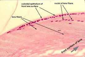
Epithelium
· Single layer of cuboidal cells
· Present only anterior to lens equator
· Basal surface towards anterior lens capsule
Nucleus and Cortex
· Oldest lens fibers (embryonic and fetal) are most central
· Lens fibers are continuously laid down peripherally
· Lens fibers originate from lens epithelial cells at the equatorial lens bow
· As lens epithelial cells de-differentiate into lens fibers, they lose all cellular organelles and nucleus
Zonules
· Support the lens
· Can be damaged by surgery, trauma, or pseudoexfoliation
· Attach from lens mid-periphery to ciliary body processes
Embryologically
· Lens originates from surface ectoderm.


Nick's Tips: Remember that these disorders are associated with systemic disease - Alport syndrome and Oculocerebralrenal syndrome.
Lenticonus / Lentiglobus
Anterior:
· Conical (lenticonus) or spherical (lentiglobus) shaped anterior lens bulge
· Has an oil-drop appearance on retroillumination during slit lamp exam.
· Bilateral cases associated with Alport Syndrome (Autosomal dominant: lenticonus, anterior polar cataract, retinal and iris neovascularization, deafness, and hemorrhagic nephritis)
· Mutations in type IV collagen are associated with lenticonus and Alport Syndrome
Posterior:
· Deformity of the posterior surface of the lens
· Oil droplet appearance as in anterior lenticonus
· Usually sporadic and unilateral
· Associated with Alport Syndrome
· Associated with Oculocerebralrenal Syndrome of Lowe (X-linked: congenital cataract, systemic acidosis, renal rickets, hypotonia)

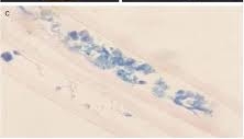
Nick's Tips: Look for history of foreign body or trauma. Prussian blue staining will show iron.
Siderosis
· Lens epithelial degeneration and necrosis
· Iron containing foreign body
· Prussian blue staining shows iron in epithelial cells